
Anatomy Of Skull
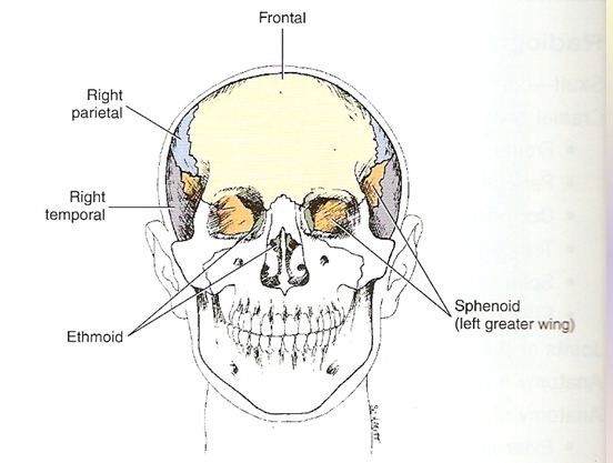
Frontal View

Lateral View
AP Axial Projection
Patient Positioning
- Depress chin, bringing OML perpendicular to IR
- Patients that unable to flex their neck, allign OML perpendicular to the IR.
- Add radiolucent support under head if needed
- Align MSP to CR and to midline of the grid / bucky
- Ensure no rotation
- IR size 24 x 30 cm
- Angle CR 30 degree caudad to OML, 36 degrees caudad to IOML
- SID 100 cm
Radiographic Image of AP Axial

Video of AP Axial
Lateral Skull
Patient Positioning
· Remove all metal, plastic or other removable
objects from head.
· Patient
erect or prone.
· Erect
may be done with dedicated head unit if
available or with erect table or other erect grid-film
• Placehead
in a true lateral position, with
side of interest closest to film
oblique body as needed for patient
comfort. (A way to check for rotation is to
palpate the external occipital protuberance posteriorly and nasion or glabella anteriorly and insure that
these two points are the same distance from the film.)
· Align midsagittal plane parallel to
film, insuring no rotation or tilt.
· To
prevent head tilting, bring interpupillary
line perpendicular to film
· Adjust
chin to bring infraorbitomeatal line perpendicular to front of cassette.
• Use 10 x 12 in. circle diaphragm or collimate to
outer margins of skull on all sides.
Radiographic Image Of Lateral Skull

Video of Lateral Projection
For more details go to HERE
No comments:
Post a Comment