Sunday 4 November 2012

Chest Radiograph

The chest x-ray is the most commonly performed diagnostic x-ray examination. A chest x-ray makes images of the heart,lungs, airways, blood vessels and the bones of the spine and chest.
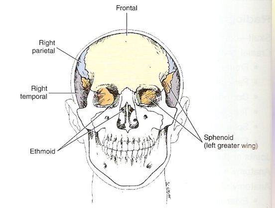


anatomy of chest


PA PROJECTION : CHEST


Pathology Demonstrated:
-pleural effusions
-pneumothorax
-atelectasis
-signs of infection
Technical Factors:
-IR size 35x43 cm (14x17 inches)
-moving or stationary grid
-110-125 kV range
-mAs 3-4
Shielding :
-lead shielding around the waist or adjustable mobile shield on a stand behind the patient
Patient position:
-patient in erect , feet spreed slighty , weight distributed equally on both feet
-chin raised , resting against IR
-hands on lower hips , palms out elbow partially flexed
-shouders rotated forward against IR to allow scapulae to move
Laterally clear of lung fields. Shoulders depressed downward to move clavicle below the apices
Part position
-alig midsaggital plane with CR and to midline of IR with equal
Margins between lateral thorax and sides of IR
-Ensure no rotation to thorax
-raise or lower CR and IR as needed to the level of T7 for average patient.
(top of IR will be 1.5 to 2 inches ,or 4 to 5 cm , above shouders on ost average patient.)
Central Ray
-CR perpendicular to IR and centered to midsaggital plane at level of T7 (7 to 8 inches or 18 to 20 cm ,below vertebra prominens or to inferior angel of scapula)
-IR centered to CR
-SID of 72 inches (180 cm)
Collimation
-collimate on four sides to area of lung field.(top borders of illuminated field should be to level of vertebra prominens and lateral borders to outer skin margins.)
 Respiration
-exposure made at end of second full inspiration 

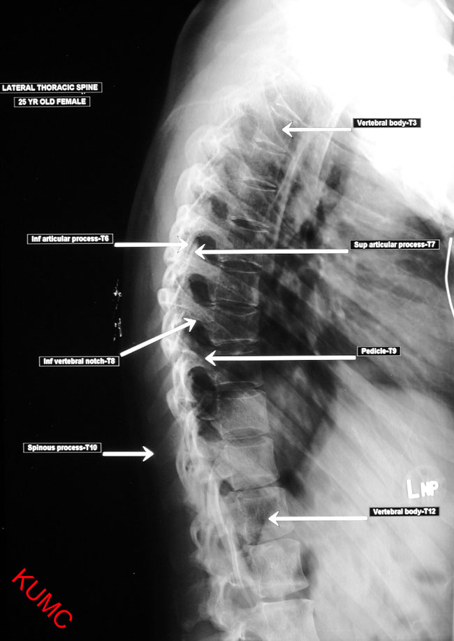
radiographic image of chest X-Ray


video of Chest radiograph positioning


for more info click right HERE


Short Form And Symbol In Medical Field


1. C/o                                    :              complaint of
2. MVA                                 :              motor vehicle accident
3. Ca                                     :              carcinoma
4. BHT                                  :              bed health ticket
5. HPT                                  :              hyperparathyroidism
6. RBC                                 :              red blood cell
7. WBC                                :              white blood cell
8. #                                       :              fracture
9. MCU                                :              micturating cyatourethrogram
  10.Ba Meal/ Swallow/Enema  :              barium meal/swallow/enema
  11.IVC                                  :              intravenous cholangeography
  12.SVC                                 :              superior vena cava
  13.CCF                                 :              congestive cardiac failure
  14.ESWL                              :              extra corporeal short wave lithotripsy
  15.CCU                                :              cardiac care unit
  16.ICU                                  :              intensive care unit
  17.cc                                     :              copies circulated
  18.UTI                                   :              urinary tract infection
  19.?                                       :              query
  20.TRO                                 :              to rule out
  21.∆                                       :              diagnosis
  22.NPC                                 :             nasopharyngeal carcinoma
  23.CSF                                  :             cerebro spinal fluid
  24.ERCP                                :             endoscopic retrograde cholongiography
  25.IVU                                   :             intravenous urography
  26.HSG                                  :             hysterosulpingography
  27.URTI                                 :             upper respiratory tract infection
  28.AMI                                  :             acute myocardial infection
  29.AVM                                 :             acute viral meningitis
  30.APG                                  :             antegrade pyelogram
  31.RPG                                  :             retrograde pyelogram
  32.DCG                                  :            dacryocystography
  33.CRHD                               :             chronic rheumatic heart disease                     
  34.IHD                                   :             ischaemic heart disease
  35.H/o                                    :             history of                                                                                               

Lumbar Positioning


Clinical Indications


Fracture
Compression
Ankylosis spondylitis
Metastases
Scoliosis
Spina bifida
•Spondylolisthesis
Spondylolysis


Anatomy of Lumbar


AP Lumbar


Lateral Lumbar


AP Lumbar positioning

The patient lies supine on the table midline aligned to the midline of the table, the hips and knees flexed 45 degrees to reduce the lumbar curve, the ASISs are equidistant from the table ensuring the midsaggital plane is at 90 degrees to the table.

Patient positioning


Central RayThe vertical central ray is centered in the midline at the level of L3 at the lower costal margin, if the sacrum and SI joints are required on the film using a 30 x 40 cm cassette the vertical central ray may be centered in the midline at the level of the iliac crests. Exposure is made on suspended expiration.


X-Ray Of AP lumbar


Video of Lumbar AP Radiography Positioning


Lateral Lumbar positioning

The patient lies on the table with their long axis in line with the long axis of the table, a line at a tangent to the third lumbar vertebra should be in line with the midline of the table. The hips and knees are flexed for stability and the hands are placed on a pillow in front of the patients face.
Ensure the line of the spinous processes are parallel to the table if not use pads to adjust the position.


Patient Positioning


Central Ray

The vertical central ray is centered in line with the highest point of the iliac crest at a point midway between the lower costal margin and the iliac crest of the raised side. Exposure is made on suspended expiration.


X-Ray Of Lateral lumbar


Video of Lumbar Lateral Radiography Positioning


for more info click right here and here


Thoracic positioning


Anatomy of Thoracic vertebra


Thoracic AP positioning

The patient lies supine on the table midline aligned to the midline of the table, the ASISs and the acromio clavicular joints are equidistant from the table ensuring the midsaggital plane is at 90 degrees to the table. The neck is extended to avoid superimposition of the mandible on the upper thoracic spine, flexion of the hips and knees may help reduce the thoracic curve.

Central RayThe vertical central ray is centered in the midline to T7 midway between the xiphisternum and the sternal angle.
Exposure is made on suspended expiration.
Exposure Factors
KvmASFFD (cm)GridFocusAECCassette
8520100YesBroadYes30 x 40 cm


X-Ray Of AP thoracic


Video of AP Thoracic Radiography Positioning


Thoracic lateral positioning

•Position : Lateral recumbent
•No rotation of patient.
•Mid coronal to centre of table.
•Place a lead sheet at the back of the patient.
•Expiration phase or long timing (sec).
•CR : perpendicular T7 at midcoronal line (8cm below the jugular notch)


Evaluation of the Image
  • ID and markers must be present and correct in the appropriate area of the film
  • Evidence of collimation on four sides equally around the centering point.
  • Optimal exposure should penetrate all the bone structures and contrast should be low enough to visualise fully the bone and soft tissue structures.
  • Limits of the examination, superiorly the upper border of C7, inferiorly the lower border of L1, laterally the medial part of the posterior ribs.
  • The intervertebral joint spaces should be visible, the spinous processes should be projected over the centers of the vertebral bodies.

X-Ray Of Lateral thoracic


Video of LateralThoracic Radiography Positioning


for more info click right here and here


Cervical x-ray


Indications
Kyphosis – abnormal convex curvature  of the thoracic spine.
Scoliosis – abnormal lateral curvature of the spine.
Spondylitis
Ankylosing spondylitis
Herniated nucleus pulposus (HNP)


Type curvature
  • cervical curve, concave forward (lordotic)
  • thoracic curve, convex forward
  • lumbar curve concave forward
  • Sacral curve , convex 


Anatomy of AP cervical


Anatomy of lateral cervical


Cervical AP positioning

•Position : oculusal plane to mastoid tip is perpendicular to IR.
•MSP is perpendicular to IR
•CR : 15º to 20º cephalic to C4 (up margin of thyroid cartilage).
FFD : 100cm

Evaluation Criteria Cervical AP

•Spinous process aligned with the midline of the cervical bodies. 

•Mandibular angles and mastoid tips are at equal distances from CV. 

•Pedicles are symmetrical lateral to the cervical bodies. 

•Dist. from the vert column to the medial ends of the clavicles are equal. 

•Intervertebral disc space open

X-Ray Of AP Cervical


Video of Cervical AP Radiography Positioning


Cervical Lateral positioning

Position : Mid coronal plane to centre of  IR.
Extend chin to prevent superimposition of mandible.
Depress shoulders.
FFD : 180 cm
CR : mid coronal  C4.

Evaluation Criteria Cervical Lateral

•Rt and Lt articular pillars and zygapophyseal joints of each cervical vertebra are superimposed. 

•Spinous process demonstrated in profile.

X-Ray Of Lateral Cervical


Video of Cervical Lateral Radiography Positioning


for more info click right here and here


SKULL

Anatomy Of Skull


Frontal View


Lateral View

AP Axial Projection

Patient Positioning 
  • Depress chin, bringing OML perpendicular to IR
  • Patients that unable to flex their neck, allign OML perpendicular to the IR.
  • Add radiolucent support under head if needed
  • Align MSP to CR and to midline of the grid / bucky
  • Ensure no rotation
  • IR size 24 x 30 cm
  • Angle CR 30 degree caudad to OML, 36 degrees caudad to IOML
  • SID 100 cm
Radiographic Image of AP Axial


Video of AP Axial


Lateral Skull

Patient Positioning

·  Remove all metal, plastic or other removable objects from head.
·  Patient erect or prone.
·  Erect may be done with dedicated head unit if available or with erect table or other erect grid-film
   Placehead in a true lateral position, with side of interest closest to film
oblique body as needed for patient comfort. (A way to check for rotation is to palpate the external occipital protuberance posteriorly and nasion or glabella anteriorly and insure that these two points are the same distance from the film.)
·  Align midsagittal plane parallel to film, insuring no rotation or tilt.
·  To prevent head tilting, bring interpupillary line perpendicular to film
·  Adjust chin to bring infraorbitomeatal line perpendicular to front of cassette.
   Use 10 x 12 in. circle diaphragm or collimate to outer margins of skull on all sides.

Radiographic Image Of Lateral Skull


Video of Lateral Projection


For more details go to HERE


Humerus AP and lateral

Indications for imaging
  • Trauma - dislocations, fractures, soft tissue calcifications
  • Arthritis survey
  • Bone pain.

anatomy of Humerus AP 


anatomy of Humerus lateral


Humerus AP positioning

Basic Patient Position

The patient stands erect AP, the position of the film is adjusted to included the shoulder and elbow joints. The patients arm is abducted approximately 20 degrees and externally rotated to bring  the distal epicondyles equidistant from the film, the patient may need to be rotated to the affected side. The humerus may need to be positioned diagonally across the film to included the full length.

Central RayThe horizontal central ray is centered midway between the skin surfaces at the midpoint of the humerus. Exposure is made on suspended expiration.

Exposure Factors

KvmASFFD (cm)GridFocusAECCassette
806100NoFineNo24x30 cm


Humerus lateral positioning


Basic Patient PositionThe patient stands erect PA, the position of the film is adjusted to included the shoulder and elbow joints, with the humerus in contact with the cassette, i.e. rotated into the anterior oblique position approximately 20 degrees. The arm is abducted approximately 35 degrees away from the trunk and the palm of the hand placed on the iliac crest to bring an imaginary line through distal humeral epicondyles at 90 degrees to the film, elbow in true lateral position.
The humerus may need to be positioned diagonally across the film to included the full length.


Central RayThe horizontal central ray is centered midway between the skin surfaces at the midpoint of the humerus. Exposure is made on suspended expiration.

Exposure Factors

KvmASFFD (cm)GridFocusAECCassette
8010100NoFineNo24x30 cm

X-Ray Of Humerus AP


X-Ray Of Humerus lateral


Video of humerus AP and Lateral Radiography Positioning


for more info click right here and here