Sunday 4 November 2012

Thoracic positioning


Anatomy of Thoracic vertebra


Thoracic AP positioning

The patient lies supine on the table midline aligned to the midline of the table, the ASISs and the acromio clavicular joints are equidistant from the table ensuring the midsaggital plane is at 90 degrees to the table. The neck is extended to avoid superimposition of the mandible on the upper thoracic spine, flexion of the hips and knees may help reduce the thoracic curve.

Central RayThe vertical central ray is centered in the midline to T7 midway between the xiphisternum and the sternal angle.
Exposure is made on suspended expiration.
Exposure Factors
KvmASFFD (cm)GridFocusAECCassette
8520100YesBroadYes30 x 40 cm


X-Ray Of AP thoracic


Video of AP Thoracic Radiography Positioning


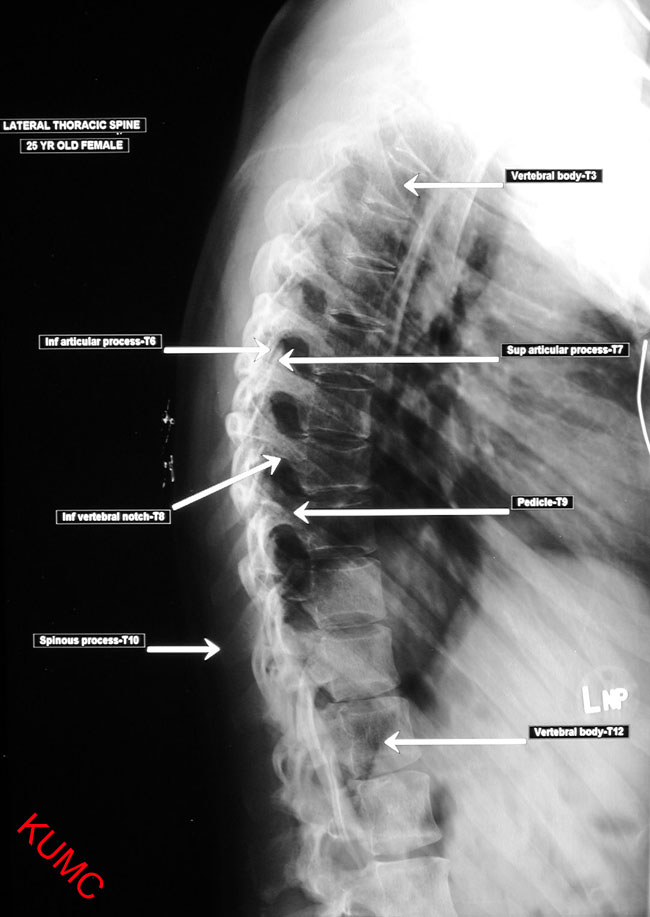
Thoracic lateral positioning

•Position : Lateral recumbent
•No rotation of patient.
•Mid coronal to centre of table.
•Place a lead sheet at the back of the patient.
•Expiration phase or long timing (sec).
•CR : perpendicular T7 at midcoronal line (8cm below the jugular notch)


Evaluation of the Image
  • ID and markers must be present and correct in the appropriate area of the film
  • Evidence of collimation on four sides equally around the centering point.
  • Optimal exposure should penetrate all the bone structures and contrast should be low enough to visualise fully the bone and soft tissue structures.
  • Limits of the examination, superiorly the upper border of C7, inferiorly the lower border of L1, laterally the medial part of the posterior ribs.
  • The intervertebral joint spaces should be visible, the spinous processes should be projected over the centers of the vertebral bodies.

X-Ray Of Lateral thoracic


Video of LateralThoracic Radiography Positioning


for more info click right here and here


No comments:

Post a Comment